Each advance in microscopic technique has provided scientists

Thông tin câu hỏi: Bộ 92 đề đọc hiểu Tiếng Anh ôn thi tốt nghiệp THPT năm 2025
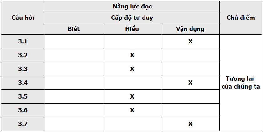
Ma trận nội dung, năng lực và cấp độ tư duy:
Read the following passage and mark the letter A, B, C, or D on your answer sheet to indicate the correct answer to each of the questions from 1 to 7.
Each advance in microscopic technique has provided scientists with new perspective, on the function of living organisms and the nature of matter itself. The invention of the visible-light microscope late in the sixteenth century introduced a previously unknown realm of single-celled plants and animals. In the twentieth century, electron microscopes have provided direct views of viruses and minuscule surface structures. Now another type of microscope, one that utilizes X rays rather than light or electrons, offers a different way of examining tiny details; it should extend human perception still farther into the natural world.
The dream of building an X-ray microscope dates to 1895; its development, however, was virtually hafted in the 1940's because the development of the electron microscope was progressing rapidly. During the 1940's electron microscopes routinely achieved resolution better than that possible with a visible-light microscope, while the performance of X-ray microscopes resisted improvement. In recent years, however, interest in X-ray microscopes has revived, largely because of advances such as the development of new sources of X-ray illumination. As a result, the brightness available today is millions of times that of X-ray tubes, which, for most of the century, were the only available sources of soft X-rays.
The new X-ray microscopes considerably improve on the resolution provided by optical microscopes. They can also be used to map the distribution of certain chemical elements. Some can form pictures in extremely short times; others hold the promise of special capabilities such as three-dimensional imaging. Unlike conventional electron microscopy, X-ray microscopy enables specimens to be kept in air and in water, which means that biological samples can be studied under conditions similar to their natural state. The illumination used, so-called soft X rays in the wavelength range of twenty to forty angstroms (an angstrom is one ten-billionth of a meter), is also sufficiently penetrating to image intact biological cells in many cases. Because of the wavelength of the X rays used, soft X-ray microscopes will never match the highest resolution possible with electron microscopes. Rather, their special properties will make possible investigations that will complement those performed with light- and electron-based instruments. (353 words)
Adapted from: https://www.scientificdiscoveries.com/microscopic-techniques
3.1. What does the passage mainly discuss?
A. The detail seen through a microscope
B. Sources of illumination for microscope
C. A new kind of microscope
D. Outdated microscopic techniques
3.2. The word "minuscule" in the first paragraph is OPPOSITE in meaning to ______.
A. circular
B. dangerous
C. complex
D. enormous
3.3. The word "enables" in paragraph 3 is CLOSEST in meaning to ______.
A. constitutes
B. specifies
C. expands
D. allows
3.4. Why did it take so long to develop the X-ray microscope?
A. Funds for research were insufficient.
B. The source of illumination was not bright enough until recently
C. Materials used to manufacture X-ray tubes were difficult to obtain.
D. X-ray microscopes were too complicated to operate.
3.5. The word "it" in paragraph 1 refers to ______.
A. a type of microscope
B. human perception
C. the natural world
D. light
3.6. According to the passage, the invention of the visible-light microscope allowed scientists to ______.
A. see viruses directly
B. develop the electron microscope later on
C. understand more about the distribution of the chemical elements
D. discover single-celled plants and animals they had never seen before
3.7. Based on the information in the passage, what can be inferred about X-ray microscopes in the future?
A. They will probably replace electron microscopes altogether.
B. They will eventually be much cheaper to produce than they are now.
C. They will provide information not available from other kinds of microscopes.
D. They will eventually chance the illumination range that they now use.
Đáp án
3.1. C
3.2. D
3.3. D
3.4. B
3.5. A
3.6. D
3.7. C
Dịch bài đọc hoàn chỉnh ra Tiếng Việt
Mỗi bước tiến trong kỹ thuật kính hiển vi đã cung cấp cho các nhà khoa học những góc nhìn mới về chức năng của các sinh vật sống và bản chất của vật chất. Sự phát minh ra kính hiển vi ánh sáng nhìn thấy vào cuối thế kỷ 16 đã giới thiệu một lĩnh vực chưa từng biết đến của các thực vật và động vật đơn bào. Vào thế kỷ 20, kính hiển vi electron đã cung cấp những cái nhìn trực tiếp về virus và các cấu trúc bề mặt nhỏ bé. Giờ đây, một loại kính hiển vi khác, sử dụng tia X thay vì ánh sáng hay electron, mang đến một cách khác để kiểm tra các chi tiết nhỏ; nó sẽ mở rộng nhận thức của con người vào thế giới tự nhiên.
Giấc mơ xây dựng một kính hiển vi tia X đã bắt đầu từ năm 1895; tuy nhiên, sự phát triển của nó gần như bị trì hoãn vào những năm 1940 do sự phát triển nhanh chóng của kính hiển vi electron. Trong những năm 1940, kính hiển vi electron thường đạt độ phân giải tốt hơn so với kính hiển vi ánh sáng nhìn thấy, trong khi hiệu suất của kính hiển vi tia X lại gặp khó khăn trong việc cải tiến. Tuy nhiên, trong những năm gần đây, sự quan tâm đến kính hiển vi tia X đã được phục hồi, chủ yếu nhờ vào những tiến bộ như việc phát triển các nguồn chiếu sáng tia X mới. Kết quả là, độ sáng có sẵn ngày nay gấp hàng triệu lần so với ống tia X, vốn là nguồn duy nhất có sẵn của tia X mềm trong hầu hết thế kỷ.
Các kính hiển vi tia X mới cải thiện đáng kể độ phân giải so với kính hiển vi quang học. Chúng cũng có thể được sử dụng để lập bản đồ phân bố của một số nguyên tố hóa học. Một số có thể tạo ra hình ảnh trong thời gian cực ngắn; những chiếc khác hứa hẹn có những khả năng đặc biệt như hình ảnh ba chiều. Khác với kính hiển vi electron thông thường, kính hiển vi tia X cho phép mẫu vật được giữ trong không khí và trong nước, điều này có nghĩa là các mẫu sinh học có thể được nghiên cứu trong điều kiện tương tự như trạng thái tự nhiên của chúng. Ánh sáng được sử dụng, gọi là tia X mềm, có bước sóng từ 20 đến 40 angstrom (một angstrom bằng một phần mười tỷ mét), cũng đủ khả năng xuyên thấu để hình ảnh hóa các tế bào sinh học nguyên vẹn trong nhiều trường hợp. Do bước sóng của tia X được sử dụng, kính hiển vi tia X mềm sẽ không bao giờ đạt được độ phân giải cao nhất như kính hiển vi electron. Thay vào đó, các đặc tính đặc biệt của chúng sẽ cho phép thực hiện các cuộc điều tra bổ sung cho các cuộc nghiên cứu được thực hiện với các dụng cụ dựa trên ánh sáng và electron.
Câu hỏi thường gặp
Xem thêm các bài cùng chuyên mục


CÔNG TY TNHH TOP EDU
Số giấy chứng nhận đăng kí kinh doanh: 0109850622, cấp ngày 09/11/2021, nơi cấp Sở Kế Hoạch và Đầu tư Thành phố Hà Nội
